Sussex Geodiversity

Sites | Treasures

Treasures

007158_detail.jpg
×

Chalk Mosasaur jaw: Mosasurus gracilis (detail), Offham nr. Lewes. (Booth Museum) (img 2)
This is part of a jaw of a marine reptile, not unlike a large monitor lizard. It would have breathed air and been a predator on fish.
Image file: 007158_detail.jpg

007158_specimen.jpg
×

Chalk Mosasaur jaw: Mosasurus gracilis, Offham nr. Lewes. (Booth Museum) (img 3)
This is part of a jaw of a marine reptile, not unlike a large monitor lizard. It would have breathed air and been a predator on fish.
Image file: 007158_specimen.jpg

007198_detail1.jpg
×

Chalk Fish: Hoplopteryx lewesiensis (detail), Upper Chalk, Southerham. (Booth Museum) (img 4)
This fish lived in the Chalk Sea 85 million years ago and was about 27cm long when fully grown.
Image file: 007198_detail1.jpg

007198_detail2.jpg
×

Chalk Fish: Hoplopteryx lewesiensis (detail), Upper Chalk, Southerham. (Booth Museum) (img 5)
This fish lived in the Chalk Sea 85 million years ago and was about 27cm long when fully grown.
Image file: 007198_detail2.jpg

007198_specimen.jpg
×

Chalk Fish: Hoplopteryx lewesiensis, Upper Chalk, Southerham. (Booth Museum) (img 6)
This fish lived in the Chalk Sea 85 million years ago and was about 27cm long when fully grown.
Image file: 007198_specimen.jpg

007750_detail_1.jpg
×

Chalk Lobster: Palaeastacus dixoni (detail) Lower Chalk, Clayton, East Sussex. (Booth Museum) (img 7)
This splendid Lobster was collected by the notable Brighton worthy Henry Willett and was first published in the scientific press in 1850!.
Image file: 007750_detail_1.jpg

007750_detail_2.jpg
×

Chalk Lobster: Palaeastacus dixoni (detail) Lower Chalk, Clayton, East Sussex. (Booth Museum) (img 8)
This splendid Lobster was collected by the notable Brighton worthy Henry Willett and was first published in the scientific press in 1850!.
Image file: 007750_detail_2.jpg

007750_specimen.jpg
×

Chalk Lobster: Palaeastacus dixoni Lower Chalk, Clayton, East Sussex. (Booth Museum) (img 9)
This splendid Lobster was collected by the notable Brighton worthy Henry Willett and was first published in the scientific press in 1850!.
Image file: 007750_specimen.jpg

007764_detail_1.jpg
×

Chalk Lobster: Enoploclytia leachii (Pincers - detail) Lower Chalk, Clayton, East Sussex. (Booth Museum) (img 10)
These pincers belong to a different type of Lobster than the last specimen.
Image file: 007764_detail_1.jpg

007764_detail_2.jpg
×

Chalk Lobster: Enoploclytia leachii (Pincers - detail) Lower Chalk, Clayton, East Sussex. (Booth Museum) (img 11)
These pincers belong to a different type of Lobster than the last specimen.
Image file: 007764_detail_2.jpg

007764_specimen.jpg
×

Chalk Lobster: Enoploclytia leachii (Pincers) Lower Chalk, Clayton, East Sussex. (Booth Museum) (img 12)
These pincers belong to a different type of Lobster than the last specimen.
Image file: 007764_specimen.jpg

007823_detail_1.jpg
×

Chalk Starfish: Metopaster sp. (detail), Upper Chalk, Kent. (Booth Museum) (img 13)
This beautiful starfish is one of many types known from the Chalk, though rarely found complete.
Image file: 007823_detail_1.jpg

007823_detail_2.jpg
×

Chalk Starfish: Metopaster sp. (detail), Upper Chalk, Kent. (Booth Museum) (img 14)
This beautiful starfish is one of many types known from the Chalk, though rarely found complete.
Image file: 007823_detail_2.jpg

007823_specimen.jpg
×

Chalk Starfish: Metopaster sp., Upper Chalk, Kent. (Booth Museum) (img 15)
This beautiful starfish is one of many types known from the Chalk, though rarely found complete.
Image file: 007823_specimen.jpg

007942_detail1.jpg
×

Chalk Crinoid: Isocrinus granosus (detail), Middle Chalk, Lewes. (Booth Museum) (img 16)
One of the best specimens known of a Chalk crinoid – a group also called sea-lilies. Imagine an upside down starfish perched on a tall stalk for an understanding of these strange creatures.
Image file: 007942_detail1.jpg

007942_detail2.jpg
×

Chalk Crinoid: Isocrinus granosus (detail), Middle Chalk, Lewes. (Booth Museum) (img 17)
One of the best specimens known of a Chalk crinoid – a group also called sea-lilies. Imagine an upside down starfish perched on a tall stalk for an understanding of these strange creatures.
Image file: 007942_detail2.jpg

007942_detail3.jpg
×

Chalk Crinoid: Isocrinus granosus (detail), Middle Chalk, Lewes. (Booth Museum) (img 18)
One of the best specimens known of a Chalk crinoid – a group also called sea-lilies. Imagine an upside down starfish perched on a tall stalk for an understanding of these strange creatures.
Image file: 007942_detail3.jpg

007942_specimen.jpg
×

Chalk Crinoid: Isocrinus granosus, Middle Chalk, Lewes. (Booth Museum) (img 19)
One of the best specimens known of a Chalk crinoid – a group also called sea-lilies. Imagine an upside down starfish perched on a tall stalk for an understanding of these strange creatures.
Image file: 007942_specimen.jpg

009057_detail.jpg
×

Chalk Bivalve: Inoceramus lamarki (detail), Upper Chalk, Brighton. (Booth Museum) (img 20)
These bivalve shellfish are common in the Chalk but are nearly always broken into small pieces. This is a rare almost complete example.
Image file: 009057_detail.jpg

009057_specimen.jpg
×

Chalk Bivalve: Inoceramus lamarki, Upper Chalk, Brighton. (Booth Museum) (img 21)
These bivalve shellfish are common in the Chalk but are nearly always broken into small pieces. This is a rare almost complete example.
Image file: 009057_specimen.jpg

011664_detail.jpg
×

Spondylus (detail) (Booth Museum) (img 22)
Spondylus spinosus was a common bivalve mollusc that lived in the Chalk Sea. It had spines which helped deter predators and supported it on soft sediments.
Image file: 011664_detail.jpg

011664_specimen.jpg
×

Spondylus (Booth Museum) (img 23)
Spondylus spinosus was a common bivalve mollusc that lived in the Chalk Sea. It had spines which helped deter predators and supported it on soft sediments.
Image file: 011664_specimen.jpg

10_Suchosaurus_1.jpg
×

Holmes Collection - Suchosaurus cultridens (Horsham Museum) (img 24)
Jaw, vertebra and scutes (bony plates) of Suchosaurus cultridens, a crocodilian. This is the first specimen listed in the Catalogue of Bax Holmes Collection, from a very young Suchosaurus found about 1837 in Horsham.
Image file: 10_Suchosaurus_1.jpg

10_Suchosaurus_2_JawDetail.jpg
×

Holmes Collection - Suchosaurus cultridens jaw bone and tooth (Horsham Museum) (img 25)
Jaw bone and tooth of Suchosaurus cultridens, a crocodilian. This is part of the first specimen listed in the Catalogue of Bax Holmes Collection, from a very young Suchosaurus found about 1837 in Horsham.
Image file: 10_Suchosaurus_2_JawDetail.jpg

10_Suchosaurus_3_ScuteDetail.jpg
×

Holmes Collection - Suchosaurus cultridens scute (Horsham Museum) (img 26)
Scute (bony plate) of Suchosaurus cultridens, a crocodilian. This is part of the first specimen listed in the Catalogue of Bax Holmes Collection, from a very young Suchosaurus found about 1837 in Horsham.
Image file: 10_Suchosaurus_3_ScuteDetail.jpg

10_Suchosaurus_3_VertebraDetail.jpg
×

Holmes Collection - Suchosaurus cultridens vertebra (Horsham Museum) (img 27)
Vertebra of Suchosaurus cultridens, a crocodilian. This is part of the first specimen listed in the Catalogue of Bax Holmes Collection, from a very young Suchosaurus found about 1837 in Horsham.
Image file: 10_Suchosaurus_3_VertebraDetail.jpg

10_Suchosaurus_3_VertebraDetail2.jpg
×

Holmes Collection - Suchosaurus cultridens vertebra (Horsham Museum) (img 28)
Vertebra of Suchosaurus cultridens, a crocodilian. This is part of the first specimen listed in the Catalogue of Bax Holmes Collection, from a very young Suchosaurus found about 1837 in Horsham.
Image file: 10_Suchosaurus_3_VertebraDetail2.jpg

11_Suchosaurus_1.jpg
×

Holmes Collection - Suchosaurus vertebra (Horsham Museum) (img 29)
Vertebra of Suchosaurus, a crocodilian. Found at Stammerham (Christ's Hospital) near Horsham.
Image file: 11_Suchosaurus_1.jpg

11_Suchosaurus_1_Detail1.jpg
×

Holmes Collection - Suchosaurus vertebra (detail) (Horsham Museum) (img 30)
Vertebra of Suchosaurus, a crocodilian. Found at Stammerham (Christ's Hospital) near Horsham.
Image file: 11_Suchosaurus_1_Detail1.jpg

11_Suchosaurus_1_Detail2.jpg
×

Holmes Collection - Suchosaurus vertebra (detail) (Horsham Museum) (img 31)
Vertebra of Suchosaurus, a crocodilian. Found at Stammerham (Christ's Hospital) near Horsham.
Image file: 11_Suchosaurus_1_Detail2.jpg

12_CrocodileTooth_1.jpg
×

Holmes Collection - crocodilian tooth (Horsham Museum) (img 32)
Tooth of crocodilian species. Found in Horsham
Image file: 12_CrocodileTooth_1.jpg

12_CrocodileTooth_1Detail.jpg
×

Holmes Collection - crocodilian tooth (detail) (Horsham Museum) (img 33)
Tooth of crocodilian species. Found in Horsham
Image file: 12_CrocodileTooth_1Detail.jpg

13_Pterodactyl_1.jpg
×

Holmes Collection - Pterodactyl wing bone (Horsham Museum) (img 34)
Wing bone of pterodactyl, a flying reptile. Specimen found at Southwater.
Image file: 13_Pterodactyl_1.jpg

13_Pterodactyl_1Detail.jpg
×

Holmes Collection - pterodactyl wing bone (detail) (Horsham Museum) (img 35)
Wing bone of pterodactyl, a flying reptile. Specimen found at Southwater.
Image file: 13_Pterodactyl_1Detail.jpg

1_Dorsal-Vertebra_1_Scale.jpg
×

Horshamosaurus - vertebra (Horsham Museum) (img 36)
Nearly complete dorsal vertebra of Horshamosaurus rudgwickensis.
Image file: 1_Dorsal-Vertebra_1_Scale.jpg

1_DorsalVertebra_1.jpg
×

Horshamosaurus - vertebra (Horsham Museum) (img 37)
Nearly complete dorsal vertebra of Horshamosaurus rudgwickensis.
Image file: 1_DorsalVertebra_1.jpg

1_DorsalVertebra_1_2.jpg
×

Horshamosaurus - vertebra (Horsham Museum) (img 38)
Nearly complete dorsal vertebra of Horshamosaurus rudgwickensis.
Image file: 1_DorsalVertebra_1_2.jpg

1_DorsalVertebra_1_Detail.jpg
×

Horshamosaurus - vertebra (detail) (Horsham Museum) (img 39)
Nearly complete dorsal vertebra of Horshamosaurus rudgwickensis.
Image file: 1_DorsalVertebra_1_Detail.jpg

1_DorsalVertebra_2.jpg
×

Horshamosaurus - vertebra (detail) (Horsham Museum) (img 40)
Nearly complete dorsal vertebra of Horshamosaurus rudgwickensis.
Image file: 1_DorsalVertebra_2.jpg

1_IguanodonFemur_1.jpg
×

Holmes Collection - Iguanodon femur (Horsham Museum) (img 41)
Distal end of large femur from the "Great Horsham Iguanodon". Found in Horsham.
Image file: 1_IguanodonFemur_1.jpg

1_IguanodonFemur_2.jpg
×

Holmes Collection - Iguanodon femur (Horsham Museum) (img 42)
Proximal end of large femur from the "Great Horsham Iguanodon". Found in Horsham.
Image file: 1_IguanodonFemur_2.jpg

2_IguanodonFemur.jpg
×

Holmes Collection - Iguanodon femur (Horsham Museum) (img 43)
Distal end of femur of Iguanodon. Found at Tower Hill, Horsham.
Image file: 2_IguanodonFemur.jpg

2_Rib_1.jpg
×

Horshamosaurus - rib (Horsham Museum) (img 44)
Part of a rib of Horshamosaurus rudgwickensis with a T-shaped cross section typical of ankylosaur ribs.
Image file: 2_Rib_1.jpg

2_Rib_1_2.jpg
×

Horshamosaurus - rib (Horsham Museum) (img 45)
Part of a rib of Horshamosaurus rudgwickensis with a T-shaped cross section typical of ankylosaur ribs.
Image file: 2_Rib_1_2.jpg

2_Rib_1_CrossSection.jpg
×

Horshamosaurus - rib (cross section) (Horsham Museum) (img 46)
Part of a rib of Horshamosaurus rudgwickensis with a T-shaped cross section typical of ankylosaur ribs.
Image file: 2_Rib_1_CrossSection.jpg

2_Rib_1_Scale.jpg
×

Horshamosaurus - rib (Horsham Museum) (img 47)
Part of a rib of Horshamosaurus rudgwickensis with a T-shaped cross section typical of ankylosaur ribs.
Image file: 2_Rib_1_Scale.jpg

3_Scapula_1.jpg
×

Horshamosaurus - scapula (Horsham Museum) (img 48)
End view of the massive and robust scapula (shoulder blade) of Horshamosaurus rudgwickensis
Image file: 3_Scapula_1.jpg

3_Scapula_1_2.jpg
×

Horshamosaurus - scapula (Horsham Museum) (img 49)
End view of the massive and robust scapula (shoulder blade) of Horshamosaurus rudgwickensis
Image file: 3_Scapula_1_2.jpg

3_Scapula_1_Detail.jpg
×

Horshamosaurus - scapula (detail) (Horsham Museum) (img 50)
Lateral view of the massive and robust scapula (shoulder blade) of Horshamosaurus rudgwickensis
Image file: 3_Scapula_1_Detail.jpg

3_Scapula_1_Scale.jpg
×

Horshamosaurus - scapula (Horsham Museum) (img 51)
Lateral view of the massive and robust scapula (shoulder blade) of Horshamosaurus rudgwickensis
Image file: 3_Scapula_1_Scale.jpg

4_DermalSpine_1.jpg
×

Horshamosaurus - dermal spine (Horsham Museum) (img 52)
Underside view of a presacral dermal spine of Horshamosaurus rudgwickensis. A piece of the dermal armour.
Image file: 4_DermalSpine_1.jpg

4_DermalSpine_1_2.jpg
×

Horshamosaurus - dermal spine (Horsham Museum) (img 53)
Underside view of a presacral dermal spine of Horshamosaurus rudgwickensis. A piece of the dermal armour.
Image file: 4_DermalSpine_1_2.jpg

4_DermalSpine_1_3.jpg
×

Horshamosaurus - dermal spine (Horsham Museum) (img 54)
Lateral view of a presacral dermal spine of Horshamosaurus rudgwickensis. A piece of the dermal armour.
Image file: 4_DermalSpine_1_3.jpg

4_DermalSpine_1_Detail.jpg
×

Horshamosaurus - dermal spine (detail) (Horsham Museum) (img 55)
Close-up of a presacral dermal spine of Horshamosaurus rudgwickensis. A piece of the dermal armour.
Image file: 4_DermalSpine_1_Detail.jpg

4_DermalSpine_1_Scale.jpg
×

Horshamosaurus - dermal spine (Horsham Museum) (img 56)
End view of a presacral dermal spine of Horshamosaurus rudgwickensis. A piece of the dermal armour.
Image file: 4_DermalSpine_1_Scale.jpg

4_Metatarsal_1.jpg
×

Holmes Collection - Iguanodon metatarsal (view from distal end) (Horsham Museum) (img 57)
Metatarsal of the "Great Horsham Iguanodon". Part of foot between toe bones and carpal bones of hind foot. Found in Horsham.
Image file: 4_Metatarsal_1.jpg

4_Metatarsal_1_2.jpg
×

Holmes Collection - Iguanodon metatarsal (lateral view) (Horsham Museum) (img 58)
Metatarsal of the "Great Horsham Iguanodon". Part of foot between toe bones and carpal bones of hind foot. Found in Horsham.
Image file: 4_Metatarsal_1_2.jpg

4_Metatarsal_1_EndDetail.jpg
×

Holmes Collection - Iguanodon metatarsal (distal end detail) (Horsham Museum) (img 59)
Metatarsal of the "Great Horsham Iguanodon". Part of foot between toe bones and carpal bones of hind foot. Found in Horsham.
Image file: 4_Metatarsal_1_EndDetail.jpg

4_Metatarsal_1_EndDetail2.jpg
×

Holmes Collection - Iguanodon metatarsal (cross section) (Horsham Museum) (img 60)
Metatarsal of the "Great Horsham Iguanodon". Part of foot between toe bones and carpal bones of hind foot. Found in Horsham.
Image file: 4_Metatarsal_1_EndDetail2.jpg

5_Metacarpal_1.jpg
×

Holmes Collection - Iguanodon metacarpal (Horsham Museum) (img 61)
Metacarpal of Iguanodon. Part of the hand, lying between the finger and wrist bones. Found near Horsham.
Image file: 5_Metacarpal_1.jpg

5_RrightTibia_1.jpg
×

Horshamosaurus - right tibia (Horsham Museum) (img 62)
Approximately two thirds of the right tibia (proximal) of Horshamosaurus rudgwickensis.
Image file: 5_RrightTibia_1.jpg

5_RrightTibia_1_2.jpg
×

Horshamosaurus - right tibia (Horsham Museum) (img 63)
Approximately two thirds of the right tibia (proximal) of Horshamosaurus rudgwickensis.
Image file: 5_RrightTibia_1_2.jpg

5_RrightTibia_1_Detail.jpg
×

Horshamosaurus - right tibia (detail) (Horsham Museum) (img 64)
Approximately two thirds of the right tibia (proximal) of Horshamosaurus rudgwickensis.
Image file: 5_RrightTibia_1_Detail.jpg

5_RrightTibia_1_Scale.jpg
×

Horshamosaurus - right tibia (Horsham Museum) (img 65)
Approximately two thirds of the right tibia (proximal) of Horshamosaurus rudgwickensis.
Image file: 5_RrightTibia_1_Scale.jpg

6_LimbBone_1.jpg
×

Horshamosaurus - limb bone (Horsham Museum) (img 66)
Unidentified limb bone from Horshamosaurus rudgwickensis.
Image file: 6_LimbBone_1.jpg

6_LimbBone_1_2.jpg
×

Horshamosaurus - limb bone (Horsham Museum) (img 67)
Unidentified limb bone from Horshamosaurus rudgwickensis.
Image file: 6_LimbBone_1_2.jpg

6_LimbBone_1_Detail.jpg
×

Horshamosaurus - limb bone (detail) (Horsham Museum) (img 68)
Unidentified limb bone from Horshamosaurus rudgwickensis.
Image file: 6_LimbBone_1_Detail.jpg

6_LimbBone_1_Scale.jpg
×

Horshamosaurus - limb bone (Horsham Museum) (img 69)
Unidentified limb bone from Horshamosaurus rudgwickensis.
Image file: 6_LimbBone_1_Scale.jpg

6_LimbBone_1_end.jpg
×

Horshamosaurus - limb bone (end view) (Horsham Museum) (img 70)
Unidentified limb bone from Horshamosaurus rudgwickensis.
Image file: 6_LimbBone_1_end.jpg

6_Metacarpal_1.jpg
×

Holmes Collection - Iguanodon metacarpal (Horsham Museum) (img 71)
Metacarpal of Iguanodon. Part of the hand, lying between the finger and wrist bones. Found near Horsham.
Image file: 6_Metacarpal_1.jpg

7_Metacarpal_1.jpg
×

Holmes Collection - Iguanodon metacarpal (Horsham Museum) (img 72)
Metacarpal of Iguanodon. Part of the hand, lying between the finger and wrist bones. Found at Stammerham (Christ's Hospital) near Horsham.
Image file: 7_Metacarpal_1.jpg

7_Rib_2.jpg
×

Holmes Collection - Iguanodon rib bone (edge view) (Horsham Museum) (img 73)
Part of rib of Iguanodon. Found in Horsham.
Image file: 7_Rib_2.jpg

7_Rib_2_2.jpg
×

Holmes Collection - Iguanodon rib bone (lateral view) (Horsham Museum) (img 74)
Part of rib of Iguanodon. Found in Horsham.
Image file: 7_Rib_2_2.jpg

7_Rib_2_EndDetail.jpg
×

Holmes Collection - Iguanodon rib bone (end detail) (Horsham Museum) (img 75)
Part of rib of Iguanodon. Found in Horsham.
Image file: 7_Rib_2_EndDetail.jpg

8_Haemapophysis_1.jpg
×

Holmes Collection - Iguanodon haemapophysis (upright view) (Horsham Museum) (img 76)
Haemapophysis from the "Great Horsham Iguanodon". Part of tail vertebra. Major blood vessels would have passed through the opening.
Image file: 8_Haemapophysis_1.jpg

8_Haemapophysis_2.jpg
×

Holmes Collection - Iguanodon haemapophysis (rotated to show base more clearly) (Horsham Museum) (img 77)
Haemapophysis from the "Great Horsham Iguanodon". Part of tail vertebra. Major blood vessels would have passed through the opening.
Image file: 8_Haemapophysis_2.jpg

8_Haemapophysis_3.jpg
×

Holmes Collection - Iguanodon haemapophysis (view from top) (Horsham Museum) (img 78)
Haemapophysis from the "Great Horsham Iguanodon". Part of tail vertebra. Major blood vessels would have passed through the opening.
Image file: 8_Haemapophysis_3.jpg

9_GoniopholisScutes_1.jpg
×

Holmes Collection - Goniopholis crassidens scutes (Horsham Museum) (img 79)
Set of scutes (bony external plates) of Goniopholis crassidens, a crocodilian. Specimens found near Horsham.
Image file: 9_GoniopholisScutes_1.jpg

9_GoniopholisScutes_2.jpg
×

Holmes Collection - Goniopholis crassidens scute (Horsham Museum) (img 80)
Single scute (bony external plate) of Goniopholis crassidens, a crocodilian. Specimen 53, found near Horsham.
Image file: 9_GoniopholisScutes_2.jpg

9_GoniopholisScutes_3.jpg
×

Holmes Collection - Goniopholis crassidens scutes (Horsham Museum) (img 81)
Two scutes (bony external plates) of Goniopholis crassidens, a crocodilian. Specimens 50 & 51, found near Horsham.
Image file: 9_GoniopholisScutes_3.jpg

9_GoniopholisScutes_4.jpg
×

Holmes Collection - Goniopholis crassidens scute (Horsham Museum) (img 82)
Single scute (bony external plate) of Goniopholis crassidens, a crocodilian. Specimen 50, found near Horsham.
Image file: 9_GoniopholisScutes_4.jpg

9_GoniopholisScutes_5.jpg
×

Holmes Collection - Goniopholis crassidens scute (detail) (Horsham Museum) (img 83)
Close-up of scute (bony external plate) of Goniopholis crassidens, a crocodilian. Middle scute of Specimen 411, found near Horsham.
Image file: 9_GoniopholisScutes_5.jpg

9_GoniopholisScutes_6.jpg
×

Holmes Collection - Goniopholis crassidens scute (Horsham Museum) (img 84)
Single scute (bony external plate) of Goniopholis crassidens, a crocodilian. Specimen 54, found near Horsham.
Image file: 9_GoniopholisScutes_6.jpg

9_GoniopholisScutes_7.jpg
×

Holmes Collection - Goniopholis crassidens scute (Horsham Museum) (img 85)
Single scute (bony external plate) of Goniopholis crassidens, a crocodilian. Specimen 52, found near Horsham.
Image file: 9_GoniopholisScutes_7.jpg

9_GoniopholisScutes_8.jpg
×

Holmes Collection - Goniopholis crassidens scutes (Horsham Museum) (img 86)
Three scutes (bony external plates) of Goniopholis crassidens, a crocodilian. Specimen 411, found near Horsham.
Image file: 9_GoniopholisScutes_8.jpg

Auroch_front.jpg
×

Auroch Skull (underside view) (Booth Museum) (img 87)
 
Image file: Auroch_front.jpg

BH_img_1.jpg
×

Polacanthus foxii spine (Bexhill Museum) (img 88)
Spine of Polacanthus foxii, a polacanthid dinosaur.
Image file: BH_img_1.jpg

BH_img_10.jpg
×

Lepidotus scales (Bexhill Museum) (img 89)
Close-up of scales at rear of Lepidotus skull.
Image file: BH_img_10.jpg

BH_img_100.jpg
×

Theropod footprint (Bexhill Museum) (img 90)
Internal cast of a dinosaur footprint, probably a theropod
Image file: BH_img_100.jpg

BH_img_101.jpg
×

Theropod footprint (Bexhill Museum) (img 91)
Internal cast of a dinosaur footprint, probably a theropod
Image file: BH_img_101.jpg

BH_img_102.jpg
×

Iguanodontid scapula (Bexhill Museum) (img 92)
Scapula (shoulder blade) of Hypselospinus, a large herbivorous Iguanodontid dinosaur
Image file: BH_img_102.jpg

BH_img_103.jpg
×

Iguanodontid scapula (Bexhill Museum) (img 93)
Scapula (shoulder blade) of Hypselospinus, a large herbivorous Iguanodontid dinosaur
Image file: BH_img_103.jpg

BH_img_104.jpg
×

Sauropod metatarsal (Bexhill Museum) (img 94)
Metatarsal (foot bone) of herbivorous sauropod dinosaur
Image file: BH_img_104.jpg

BH_img_105.jpg
×

Sauropod metatarsal (Bexhill Museum) (img 95)
Metatarsal (foot bone) of herbivorous sauropod dinosaur.
Image file: BH_img_105.jpg

BH_img_106.jpg
×

Sauropod metatarsal (Bexhill Museum) (img 96)
Close up of surface of the metatarsal (foot bone) of a sauropod dinosaur, shown in previous images.
Image file: BH_img_106.jpg

BH_img_107.jpg
×

Sauropod metatarsal (Bexhill Museum) (img 97)
Close up of surface of the metatarsal (foot bone) of a sauropod dinosaur, shown in previous images.
Image file: BH_img_107.jpg

BH_img_108.jpg
×

Hypsilospinus dorsal spine (Bexhill Museum) (img 98)
Neural processes and dorsal spine of Hypsilospinus, a large iguanodontid dinosaur
Image file: BH_img_108.jpg

BH_img_109.jpg
×

Hypsilospinus dorsal spine (Bexhill Museum) (img 99)
Neural processes and dorsal spine of Hypsilospinus, a large iguanodontid dinosaur
Image file: BH_img_109.jpg

BH_img_11.jpg
×

Lepidotus scales (Bexhill Museum) (img 100)
Scales of Lepidotus, a neopterygian ray-finned fish.
Image file: BH_img_11.jpg

BH_img_110.jpg
×

Hypsilospinus dorsal spine (Bexhill Museum) (img 101)
Neural processes and dorsal spine of Hypsilospinus, a large iguanodontid dinosaur
Image file: BH_img_110.jpg

BH_img_111.jpg
×

Hypsilospinus dorsal spine (Bexhill Museum) (img 102)
Neural processes and dorsal spine of Hypsilospinus, a large iguanodontid dinosaur
Image file: BH_img_111.jpg

BH_img_112.jpg
×

Hypsilospinus dorsal spine (Bexhill Museum) (img 103)
Neural processes and dorsal spine of Hypsilospinus, a large iguanodontid dinosaur
Image file: BH_img_112.jpg

BH_img_12.jpg
×

Lepidotus scales (Bexhill Museum) (img 104)
Close-up of scales of Lepidotus
Image file: BH_img_12.jpg

BH_img_13.jpg
×

Lepidotus scales (Bexhill Museum) (img 105)
Close-up of scales of Lepidotus
Image file: BH_img_13.jpg

BH_img_14.jpg
×

Lepidotus scales (Bexhill Museum) (img 106)
Close-up of scales of Lepidotus
Image file: BH_img_14.jpg

BH_img_15.jpg
×

Lepidotus vertebrae (Bexhill Museum) (img 107)
In situ vertebrae of Lepidotus, a neopterygian ray-finned fish. Also shows lateral processes.
Image file: BH_img_15.jpg

BH_img_16.jpg
×

Lepidotus vertebrae (Bexhill Museum) (img 108)
Close-up of vertebrae and lateral processes of Lepidotus.
Image file: BH_img_16.jpg

BH_img_17.jpg
×

Lepidotus vertebrae (Bexhill Museum) (img 109)
Close-up of vertebrae of Lepidotus.
Image file: BH_img_17.jpg

BH_img_18.jpg
×

Williamsonia (Bexhill Museum) (img 110)
Flower-like reproductive structure of Williamsonia, a bennettitalean (cycadeoid) plant. Wadhurst Clay, Ashdown Formation.
Image file: BH_img_18.jpg

BH_img_19.jpg
×

Hybodus spine (Bexhill Museum) (img 111)
Spine of Hybodus, a small freshwater shark which had a spine in front of each dorsal fin
Image file: BH_img_19.jpg

BH_img_2.jpg
×

Polacanthus foxii spine (Bexhill Museum) (img 112)
Spine of Polacanthus foxii, a polacanthid dinosaur.
Image file: BH_img_2.jpg

BH_img_20.jpg
×

Hybodus spine (Bexhill Museum) (img 113)
Close-up of tip of spine of Hybodus
Image file: BH_img_20.jpg

BH_img_21.jpg
×

Hybodus spine (Bexhill Museum) (img 114)
Close-up of tip of spine of Hybodus
Image file: BH_img_21.jpg

BH_img_22.jpg
×

Hybodus spine (Bexhill Museum) (img 115)
Close-up of tip of spine of Hybodus
Image file: BH_img_22.jpg

BH_img_23.jpg
×

Hybodus spine (Bexhill Museum) (img 116)
Close-up of centre of spine of Hybodus
Image file: BH_img_23.jpg

BH_img_24.jpg
×

Hybodus spine (Bexhill Museum) (img 117)
Head spine of Hybodus, a small freshwater shark
Image file: BH_img_24.jpg

BH_img_25.jpg
×

Hybodus spine (Bexhill Museum) (img 118)
Head spine of Hybodus, a small freshwater shark
Image file: BH_img_25.jpg

BH_img_26.jpg
×

Hybodus spine (Bexhill Museum) (img 119)
Head spine of Hybodus, a small freshwater shark
Image file: BH_img_26.jpg

BH_img_27.jpg
×

Hybodus spine (Bexhill Museum) (img 120)
Head spine of Hybodus, a small freshwater shark
Image file: BH_img_27.jpg

BH_img_28.jpg
×

Hybodus spine (Bexhill Museum) (img 121)
Head spine of Hybodus, a small freshwater shark
Image file: BH_img_28.jpg

BH_img_29.jpg
×

Hybodus tooth (Bexhill Museum) (img 122)
Outer side of tooth of Hybodus, a small freshwater shark
Image file: BH_img_29.jpg

BH_img_3.jpg
×

Iguanodontid vertebra (Bexhill Museum) (img 123)
Cervical vertebra of iguanodontid herbivorous dinosaur
Image file: BH_img_3.jpg

BH_img_30.jpg
×

Hybodus tooth (Bexhill Museum) (img 124)
Inner side of tooth of Hybodus, a small freshwater shark
Image file: BH_img_30.jpg

BH_img_31.jpg
×

Hybodus tooth (Bexhill Museum) (img 125)
View of inner side of tooth of Hybodus shown in previous image
Image file: BH_img_31.jpg

BH_img_32.jpg
×

Hybodus tooth (Bexhill Museum) (img 126)
Outer side of tooth of Hybodus shown in previous image
Image file: BH_img_32.jpg

BH_img_33.jpg
×

Lepidotus fin (Bexhill Museum) (img 127)
Bone from fin of Lepidotus, a neopterygian ray-finned fish
Image file: BH_img_33.jpg

BH_img_34.jpg
×

Lepidotus fin (Bexhill Museum) (img 128)
Bone from fin of Lepidotus, a neopterygian ray-finned fish
Image file: BH_img_34.jpg

BH_img_35.jpg
×

Lepidotus fin (Bexhill Museum) (img 129)
Close-up of end of bone from fin of Lepidotus
Image file: BH_img_35.jpg

BH_img_36.jpg
×

Ornithocheirus tooth (Bexhill Museum) (img 130)
Tooth of Ornithocheirus, a pterosaur (flying reptile)
Image file: BH_img_36.jpg

BH_img_37.jpg
×

Ornithocheirus tooth (Bexhill Museum) (img 131)
Tooth of Ornithocheirus, a pterosaur (flying reptile)
Image file: BH_img_37.jpg

BH_img_38.jpg
×

Ornithocheirus tooth (Bexhill Museum) (img 132)
Close-up of tip of tooth from Ornithocheirus, a pterosaur
Image file: BH_img_38.jpg

BH_img_39.jpg
×

Turtle carapace (Bexhill Museum) (img 133)
Part of carapace of a turtle. Bones supporting the shell.
Image file: BH_img_39.jpg

BH_img_4.5.jpg
×

Turtle carapace (Bexhill Museum) (img 134)
Part of carapace of a turtle. Bones supporting the shell.
Image file: BH_img_4.5.jpg

BH_img_4.jpg
×

Iguanodontid vertebra (Bexhill Museum) (img 135)
Cervical vertebra of iguanodontid herbivorous dinosaur
Image file: BH_img_4.jpg

BH_img_40.jpg
×

Turtle carapace (Bexhill Museum) (img 136)
Close-up of bones from carapace of turtle. Specimen shown in previous image.
Image file: BH_img_40.jpg

BH_img_41.jpg
×

Turtle carapace (Bexhill Museum) (img 137)
Part of carapace of a turtle. Bones supporting the shell. Underside of specimen in earlier image.
Image file: BH_img_41.jpg

BH_img_42.jpg
×

Turtle carapace (Bexhill Museum) (img 138)
Close-up of bones from carapace of turtle. Specimen shown in earlier image
Image file: BH_img_42.jpg

BH_img_43.jpg
×

Aigialosaurus jawbone (Bexhill Museum) (img 139)
Jawbone and teeth of Aigialosaurus, a small marine reptile, precursor of the large mosasaurs
Image file: BH_img_43.jpg

BH_img_44.jpg
×

Aigialosaurus jawbone (Bexhill Museum) (img 140)
Jawbone and teeth of Aigialosaurus, a small marine reptile, precursor of the large mosasaurs
Image file: BH_img_44.jpg

BH_img_45.jpg
×

Aigialosaurus teeth (Bexhill Museum) (img 141)
Close-up of teeth of Aigialosaurus
Image file: BH_img_45.jpg

BH_img_46.jpg
×

Allosaurid tooth (Bexhill Museum) (img 142)
Tooth of an Allosaurid carnivorous dinosaur
Image file: BH_img_46.jpg

BH_img_47.jpg
×

Allosaurid tooth (Bexhill Museum) (img 143)
Another view of the tooth of an Allosaurid carnivorous dinosaur
Image file: BH_img_47.jpg

BH_img_48.jpg
×

Allosaurid tooth (Bexhill Museum) (img 144)
Closer view of the tooth of an Allosaurid carnivorous dinosaur
Image file: BH_img_48.jpg

BH_img_49.jpg
×

Baryonyx tooth (Bexhill Museum) (img 145)
Tooth of Baryonyx, a large carnivorous dinosaur which specialised in eating fish. Tooth is ribbed like a crocodile tooth but has serrations unlike crocodiles.
Image file: BH_img_49.jpg

BH_img_4point5.jpg
×

Iguanodontid vertebra (Bexhill Museum) (img 146)
Cervical vertebra of iguanodontid herbivorous dinosaur
Image file: BH_img_4point5.jpg

BH_img_5.jpg
×

Iguanodontid vertebra (Bexhill Museum) (img 147)
Cervical vertebra of iguanodontid dinosaur. Close-up of cancellus bone tissue at the core of the bone.
Image file: BH_img_5.jpg

BH_img_50.jpg
×

Baryonyx tooth (Bexhill Museum) (img 148)
Tooth of Baryonyx, a large carnivorous dinosaur which specialised in eating fish. Tooth is ribbed like a crocodile tooth but has serrations unlike crocodiles.
Image file: BH_img_50.jpg

BH_img_51.jpg
×

Baryonyx tooth (Bexhill Museum) (img 149)
Tooth of Baryonyx, a large carnivorous dinosaur which specialised in eating fish. Tooth is ribbed like a crocodile tooth but has serrations unlike crocodiles.
Image file: BH_img_51.jpg

BH_img_52.jpg
×

Polacanthus tooth (Bexhill Museum) (img 150)
Tooth of Polacanthus, an ankylosaurid herbivorous dinosaur. From Ashdown Formation, Wadhurst Clay.
Image file: BH_img_52.jpg

BH_img_53.jpg
×

Polacanthus tooth (Bexhill Museum) (img 151)
Close-up of tooth of Polacanthus.
Image file: BH_img_53.jpg

BH_img_54.jpg
×

Dromaeosaurid tooth (Bexhill Museum) (img 152)
Tooth of Dromaeosaurid dinosaur showing a serrated edge. A bird like dinosaur which may have had feathers.
Image file: BH_img_54.jpg

BH_img_55.jpg
×

Dromaeosaurid tooth (Bexhill Museum) (img 153)
Tooth of Dromaeosaurid dinosaur showing serrated edges. A bird like dinosaur which may have had feathers.
Image file: BH_img_55.jpg

BH_img_56.jpg
×

Dromaeosaurid tooth (Bexhill Museum) (img 154)
Tooth of Dromaeosaurid dinosaur showing serrated edges. A bird like dinosaur which may have had feathers.
Image file: BH_img_56.jpg

BH_img_57.jpg
×

Dromaeosaurid tooth (Bexhill Museum) (img 155)
Tooth of Dromaeosaurid dinosaur showing serrated edges. A bird like dinosaur which may have had feathers.
Image file: BH_img_57.jpg

BH_img_58.jpg
×

Unio (Bexhill Museum) (img 156)
Shell of Unio, a freshwater mussel
Image file: BH_img_58.jpg

BH_img_59.jpg
×

Unio (Bexhill Museum) (img 157)
Lateral view of shell of Unio, a freshwater mussel
Image file: BH_img_59.jpg

BH_img_6.jpg
×

Lepidotus skull (Bexhill Museum) (img 158)
Frontal view of skull of Lepidotus with plates and teeth in situ. Lepidotus was a neopterygian ray-finned fish.
Image file: BH_img_6.jpg

BH_img_60.jpg
×

Wealden amber (Bexhill Museum) (img 159)
Exceptionally rare amber from Wealden Group associated with fossil fir tree. No insects were found in this specimen.
Image file: BH_img_60.jpg

BH_img_61.jpg
×

Wealden amber (Bexhill Museum) (img 160)
Exceptionally rare amber from Wealden Group associated with fossil fir tree. No insects were found in this specimen.
Image file: BH_img_61.jpg

BH_img_62.jpg
×

Iguanodon teeth (Bexhill Museum) (img 161)
Two teeth from Iguanodon, a large herbivorous dinosaur. Shows grinding surfaces.
Image file: BH_img_62.jpg

BH_img_63.jpg
×

Iguanodon tooth (Bexhill Museum) (img 162)
Close-up of the smaller Iguanodon tooth shown in previous image
Image file: BH_img_63.jpg

BH_img_64.jpg
×

Iguanodon tooth (Bexhill Museum) (img 163)
Close-up of the larger Iguanodon tooth shown in earlier image
Image file: BH_img_64.jpg

BH_img_65.jpg
×

Coprolite (Bexhill Museum) (img 164)
Coprolite, fossil dung of crocodile or dinosaur. Contains fish scales.
Image file: BH_img_65.jpg

BH_img_66.jpg
×

Coprolite (Bexhill Museum) (img 165)
Coprolite, fossil dung of crocodile or dinosaur. Contains fish scales.
Image file: BH_img_66.jpg

BH_img_67.jpg
×

Coprolite (Bexhill Museum) (img 166)
Close-up of coprolite showing fish scales
Image file: BH_img_67.jpg

BH_img_69.jpg
×

Crocodile scute (Bexhill Museum) (img 167)
Crocodile scute, possibly from skull
Image file: BH_img_69.jpg

BH_img_7.jpg
×

Lepidotus skull (Bexhill Museum) (img 168)
Lateral view of skull of Lepidotus with plates and teeth in situ. Lepidotus was a neopterygian ray-finned fish.
Image file: BH_img_7.jpg

BH_img_70.jpg
×

Crocodile scute (Bexhill Museum) (img 169)
Underside of crocodile scute shown in previous image. Possibly from skull.
Image file: BH_img_70.jpg

BH_img_71.jpg
×

Crocodile scute (Bexhill Museum) (img 170)
Crocodile scute, from body
Image file: BH_img_71.jpg

BH_img_72.jpg
×

Crocodile scute (Bexhill Museum) (img 171)
Close-up of crocodile scute shown in earlier image. Possibly from skull.
Image file: BH_img_72.jpg

BH_img_73.jpg
×

Crocodile scute (Bexhill Museum) (img 172)
Close-up of crocodile scute shown in earlier image. Possibly from skull.
Image file: BH_img_73.jpg

BH_img_74.jpg
×

Bucklandia (Bexhill Museum) (img 173)
Stem of Bucklandia, a bennettitalean (cycadeoid) plant
Image file: BH_img_74.jpg

BH_img_75.jpg
×

Bucklandia (Bexhill Museum) (img 174)
Close-up of stem of Bucklandia
Image file: BH_img_75.jpg

BH_img_76.jpg
×

Tempskya (Bexhill Museum) (img 175)
Close-up of interior of stem of Tempskya.
Image file: BH_img_76.jpg

BH_img_77.jpg
×

Tempskya (Bexhill Museum) (img 176)
Close-up of interior of stem of Tempskya.
Image file: BH_img_77.jpg

BH_img_78.jpg
×

Tempskya (Bexhill Museum) (img 177)
Close-up of interior of stem of Tempskya.
Image file: BH_img_78.jpg

BH_img_79.jpg
×

Tempskya (Bexhill Museum) (img 178)
Interior of stem of Tempskya, a tree-like fern
Image file: BH_img_79.jpg

BH_img_8.jpg
×

Lepidotus teeth (Bexhill Museum) (img 179)
Close-up of mouth of Lepidotus showing teeth in situ.
Image file: BH_img_8.jpg

BH_img_80.jpg
×

Lepidotus teeth (Bexhill Museum) (img 180)
Close-up of mouth of Lepidotus showing teeth in situ.
Image file: BH_img_80.jpg

BH_img_81.jpg
×

Maniraptoran vertebra (Bexhill Museum) (img 181)
Neck bone of Maniraptoran dinosaur found at Ashdown Bickworks, Bexhill. Very bird-like and one of the world's smallest dinosaurs.
Image file: BH_img_81.jpg

BH_img_82.jpg
×

Maniraptoran vertebra (Bexhill Museum) (img 182)
Neck bone of Maniraptoran dinosaur found at Ashdown Bickworks, Bexhill. Very bird-like and one of the world's smallest dinosaurs.
Image file: BH_img_82.jpg

BH_img_83.jpg
×

Maniraptoran vertebra (Bexhill Museum) (img 183)
Neck bone of Maniraptoran dinosaur found at Ashdown Bickworks, Bexhill. Very bird-like and one of the world's smallest dinosaurs.
Image file: BH_img_83.jpg

BH_img_85.jpg
×

Maniraptoran vertebra (Bexhill Museum) (img 184)
Neck bone of Maniraptoran dinosaur found at Ashdown Bickworks, Bexhill. Very bird-like and one of the world's smallest dinosaurs.
Image file: BH_img_85.jpg

BH_img_86.jpg
×

Sauropod tail bone (Bexhill Museum) (img 185)
Tail bone from a sauropod herbivorous dinosaur, probably a Titanosaur. The neural arch in these sauropods only extended half way along the tail.
Image file: BH_img_86.jpg

BH_img_87.jpg
×

Sauropod tail bone (Bexhill Museum) (img 186)
Tail bone from a sauropod herbivorous dinosaur, probably a Titanosaur. The neural arch in these sauropods only extended half way along the tail.
Image file: BH_img_87.jpg

BH_img_88.jpg
×

Sauropod tail bone (Bexhill Museum) (img 187)
Close-up of tail bone of sauropod dinosaur
Image file: BH_img_88.jpg

BH_img_89.jpg
×

Sauropod tail bone (Bexhill Museum) (img 188)
Close-up cross section of tail bone of sauropod dinosaur
Image file: BH_img_89.jpg

BH_img_9.jpg
×

Lepidotus teeth (Bexhill Museum) (img 189)
Close-up of mouth of Lepidotus showing teeth in situ and skeletons of bryozoa which grew on the jaws after death.
Image file: BH_img_9.jpg

BH_img_90.jpg
×

Iguanodon jawbone (Bexhill Museum) (img 190)
Jawbone and teeth of Iguanodon, a large herbivorous dinosaur. Replacement teeth visible.
Image file: BH_img_90.jpg

BH_img_91.jpg
×

Iguanodon jawbone (Bexhill Museum) (img 191)
Close-up of replacement tooth in jaw of Iguanodon
Image file: BH_img_91.jpg

BH_img_92.jpg
×

Iguanodon jawbone (Bexhill Museum) (img 192)
Close-up of replacement tooth in jaw of Iguanodon
Image file: BH_img_92.jpg

BH_img_94.jpg
×

Iguanodon jawbone (Bexhill Museum) (img 193)
Close-up of jaw bone of Iguanodon
Image file: BH_img_94.jpg

BH_img_95.jpg
×

Iguanodon shoulder bone (Bexhill Museum) (img 194)
Part of the pectoral (shoulder) girdle of an Iguanodontid dinosaur, a large herbivorous dinosaur
Image file: BH_img_95.jpg

BH_img_96.jpg
×

Iguanodon shoulder bone (Bexhill Museum) (img 195)
Close-up of pectoral (shoulder) girdle of Iguanodontid dinosaur, shown in previous images
Image file: BH_img_96.jpg

BH_img_97.jpg
×

Iguanodon shoulder bone (Bexhill Museum) (img 196)
Close-up of pectoral (shoulder) girdle of Iguanodontid dinosaur, shown in previous images
Image file: BH_img_97.jpg

BH_img_98.jpg
×

Iguanodon shoulder bone (Bexhill Museum) (img 197)
Part of the pectoral (shoulder) girdle of an Iguanodontid dinosaur, a large herbivorous dinosaur
Image file: BH_img_98.jpg

BH_img_99.jpg
×

Theropod footprint (Bexhill Museum) (img 198)
Internal cast of a dinosaur footprint, probably a theropod
Image file: BH_img_99.jpg

Dragonfly_1.jpg
×

Dragonflies - Libellulium zdrzaleki (Horsham Museum) (img 199)
Part and counterpart of hindwing of petalurid dragonfly Libellulium zdrzaleki.
Image file: Dragonfly_1.jpg

Dragonfly_1Counterpart.jpg
×

Dragonflies - Libellulium zdrzaleki (Horsham Museum) (img 200)
Counterpart of hindwing of petalurid dragonfly Libellulium zdrzaleki.
Image file: Dragonfly_1Counterpart.jpg

Dragonfly_1Part.jpg
×

Dragonflies - Libellulium zdrzaleki (Horsham Museum) (img 201)
Part of hindwing of petalurid dragonfly Libellulium zdrzaleki.
Image file: Dragonfly_1Part.jpg

Dragonfly_2.jpg
×

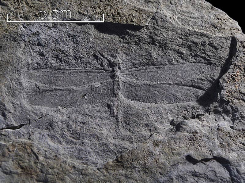
Dragonflies - Libellulium standingae (Horsham Museum) (img 202)
Hindwing of petalurid dragonfly Libellulium standingae.
Image file: Dragonfly_2.jpg

Dragonfly_2Detail1.jpg
×

Dragonflies - Libellulium standingae (Horsham Museum) (img 203)
Hindwing of petalurid dragonfly Libellulium standingae.
Image file: Dragonfly_2Detail1.jpg

Dragonfly_2WingBase.jpg
×

Dragonflies - Libellulium standingae (Horsham Museum) (img 204)
Base of hindwing of petalurid dragonfly Libellulium standingae.
Image file: Dragonfly_2WingBase.jpg

Dragonfly_2WingBase2.jpg
×

Dragonflies - Libellulium standingae (Horsham Museum) (img 205)
Base of hindwing of petalurid dragonfly Libellulium standingae.
Image file: Dragonfly_2WingBase2.jpg

Dragonfly_2WingTip.jpg
×

Dragonflies - Libellulium standingae (Horsham Museum) (img 206)
Wing-tip of petalurid dragonfly Libellulium standingae.
Image file: Dragonfly_2WingTip.jpg

Dragonfly_2_Scale.jpg
×

Dragonflies - Libellulium standingae (Horsham Museum) (img 207)
Wing-tip of petalurid dragonfly Libellulium standingae.
Image file: Dragonfly_2_Scale.jpg

Echinoderm_detail.jpg
×

Dragonflies - Libellulium standingae (Horsham Museum) (img 208)
Wing-tip of petalurid dragonfly Libellulium standingae.
Image file: Echinoderm_detail.jpg

Echinoderm_specimen.jpg
×

Dragonflies - Libellulium standingae (Horsham Museum) (img 209)
Wing-tip of petalurid dragonfly Libellulium standingae.
Image file: Echinoderm_specimen.jpg

Fish.jpg
×

Ctenothrissa radians Upper Chalk, East Sussex. (Booth Museum) (img 210)
Found by Charles Potter in the Lewes area in the 19th century this is a rare complete example of this beautiful scaly fish.
Image file: Fish.jpg

Fish_016957.jpg
×

Ctenothrissa radians Upper Chalk, East Sussex. (Booth Museum) (img 211)
Found by Charles Potter in the Lewes area in the 19th century this is a rare complete example of this beautiful scaly fish.
Image file: Fish_016957.jpg

Fish_016957_detail_1.jpg
×

Ctenothrissa radians Upper Chalk, East Sussex. (Booth Museum) (img 212)
Found by Charles Potter in the Lewes area in the 19th century this is a rare complete example of this beautiful scaly fish.
Image file: Fish_016957_detail_1.jpg

Fish_016957_detail_2.jpg
×

Ctenothrissa radians Upper Chalk, East Sussex. (Booth Museum) (img 213)
Found by Charles Potter in the Lewes area in the 19th century this is a rare complete example of this beautiful scaly fish.
Image file: Fish_016957_detail_2.jpg

Fish_016957_detail_3.jpg
×

Ctenothrissa radians Upper Chalk, East Sussex. (Booth Museum) (img 214)
Found by Charles Potter in the Lewes area in the 19th century this is a rare complete example of this beautiful scaly fish.
Image file: Fish_016957_detail_3.jpg

Fish_detail_1.jpg
×

Ctenothrissa radians (detail) Upper Chalk, East Sussex. (Booth Museum) (img 215)
Found by Charles Potter in the Lewes area in the 19th century this is a rare complete example of this beautiful scaly fish.
Image file: Fish_detail_1.jpg

Fish_detail_2.jpg
×

Ctenothrissa radians (detail) Upper Chalk, East Sussex. (Booth Museum) (img 216)
Found by Charles Potter in the Lewes area in the 19th century this is a rare complete example of this beautiful scaly fish.
Image file: Fish_detail_2.jpg

Fish_detail_3.jpg
×

Ctenothrissa radians (detail) Upper Chalk, East Sussex. (Booth Museum) (img 217)
Found by Charles Potter in the Lewes area in the 19th century this is a rare complete example of this beautiful scaly fish.
Image file: Fish_detail_3.jpg

HybodusSketch.jpg
×

Hybodus (Bexhill Museum) (img 218)
Sketch
Image file: HybodusSketch.jpg

IMGP3434edit.jpg
×

Hybodus (Bexhill Museum) (img 219)
Sketch
Image file: IMGP3434edit.jpg

IMGP3439edit.jpg
×

Hybodus (Bexhill Museum) (img 220)
Sketch
Image file: IMGP3439edit.jpg

IMGP3665edit.jpg
×

Hybodus (Bexhill Museum) (img 221)
Sketch
Image file: IMGP3665edit.jpg

IMGP3765edit.jpg
×

Hybodus (Bexhill Museum) (img 222)
Sketch
Image file: IMGP3765edit.jpg

IMGP8780.jpg
×

Crocodile skull from Cuckfield. (from above) (Booth Museum) (img 223)
This splendid skull is from about 140 million years ago and belonged to an early crocodile that lived in Sussex together with dinosaurs like Iguanodon. Unfortunately its lower jaw and most teeth are missing.
Image file: IMGP8780.jpg

IMGP8783.jpg
×

Crocodile skull from Cuckfield. (from below) (Booth Museum) (img 224)
This splendid skull is from about 140 million years ago and belonged to an early crocodile that lived in Sussex together with dinosaurs like Iguanodon. Unfortunately its lower jaw and most teeth are missing.
Image file: IMGP8783.jpg

IMGP8804.jpg
×

Auroch Skull (top view) (Booth Museum) (img 225)
 
Image file: IMGP8804.jpg

IMGP8811.jpg
×

Iguanodon toe (Booth Museum) (img 226)
Iguanodon is the dinosaur most famously connected with Sussex since it was here where its teeth were first found in 1822 by Gideon Mantell, the Lewes doctor. This toe - or metatarsal is the fourth bone up from the claw bone.
Image file: IMGP8811.jpg

IMGP8815.jpg
×

Iguanodon toe (detail) (Booth Museum) (img 227)
Iguanodon is the dinosaur most famously connected with Sussex since it was here where its teeth were first found in 1822 by Gideon Mantell, the Lewes doctor. This toe - or metatarsal is the fourth bone up from the claw bone.
Image file: IMGP8815.jpg

IMGP8831.jpg
×

Iguanodon footprint cast (Booth Museum) (img 228)
This is a natural cast of an Iguanodon footprint from the beach at Bexhill. They are occasionally uncovered at low tides.
Image file: IMGP8831.jpg

IMGP8842.jpg
×

Iguanodon femur (Booth Museum) (img 229)
Part of the thigh bone of an Iguanodon showing just how massive its legs were.
Image file: IMGP8842.jpg

IMGP8845.jpg
×

Iguanodon femur (end view) (Booth Museum) (img 230)
Part of the thigh bone of an Iguanodon showing just how massive its legs were.
Image file: IMGP8845.jpg

IMGP8849.jpg
×

Iguanodon vertebra (Booth Museum) (img 231)
A bone from the tail of a young dinosaur - probably Iguanodon.
Image file: IMGP8849.jpg

IMGP8855.jpg
×

Iguanodon vertebra (Booth Museum) (img 232)
A bone from the tail of a young dinosaur - probably Iguanodon.
Image file: IMGP8855.jpg

IMGP8860.jpg
×

Iguanodon small toe (Booth Museum) (img 233)
A toe from a young dinosaur.
Image file: IMGP8860.jpg

IMGP8865.jpg
×

Iguanodon pubis (Booth Museum) (img 234)
When this was first found in the early 19th century no-one was sure what bone it was. First identified as a shoulder blade it turned out to be part of the pelvis of a young Iguanodon.
Image file: IMGP8865.jpg

IMGP8872.jpg
×

Crocodile snout (lower) (Booth Museum) (img 235)
This snout preserves some of the teeth which the crocodile would have used to catch the large fish which swam in the rivers and lakes 140 million years ago.
Image file: IMGP8872.jpg

IMGP8878.jpg
×

Crocodile snout (detail) (Booth Museum) (img 236)
This snout preserves some of the teeth which the crocodile would have used to catch the large fish which swam in the rivers and lakes 140 million years ago.
Image file: IMGP8878.jpg

IMGP8884.jpg
×

Crocodile snout (Booth Museum) (img 237)
This snout preserves some of the teeth which the crocodile would have used to catch the large fish which swam in the rivers and lakes 140 million years ago.
Image file: IMGP8884.jpg

IMGP8890.jpg
×

Insects: Odonata (Booth Museum) (img 238)
The hot moist conditions of the monsoonal Weald, 140 million years ago would have suited insects of all kinds. If you know where to look insect fossils are quite common in rocks of Sussex and Surrey and include dragonflies, midges, ants, daddy long legs, wasps and cockroaches.
Image file: IMGP8890.jpg
IMGP8895.jpg
×

Insects: Odonata (Booth Museum) (img 239)
The hot moist conditions of the monsoonal Weald, 140 million years ago would have suited insects of all kinds. If you know where to look insect fossils are quite common in rocks of Sussex and Surrey and include dragonflies, midges, ants, daddy long legs, wasps and cockroaches.
Image file: IMGP8895.jpg

IMGP8910.jpg
×

Insects: Odonata (Booth Museum) (img 240)
The hot moist conditions of the monsoonal Weald, 140 million years ago would have suited insects of all kinds. If you know where to look insect fossils are quite common in rocks of Sussex and Surrey and include dragonflies, midges, ants, daddy long legs, wasps and cockroaches.
Image file: IMGP8910.jpg

IMGP8914.jpg
×

Insects: Odonata (Booth Museum) (img 241)
The hot moist conditions of the monsoonal Weald, 140 million years ago would have suited insects of all kinds. If you know where to look insect fossils are quite common in rocks of Sussex and Surrey and include dragonflies, midges, ants, daddy long legs, wasps and cockroaches.
Image file: IMGP8914.jpg

IMGP8920.jpg
×

Insects: Odonata (Booth Museum) (img 242)
The hot moist conditions of the monsoonal Weald, 140 million years ago would have suited insects of all kinds. If you know where to look insect fossils are quite common in rocks of Sussex and Surrey and include dragonflies, midges, ants, daddy long legs, wasps and cockroaches.
Image file: IMGP8920.jpg

IMGP8927.jpg
×

Insects: Odonata (Booth Museum) (img 243)
The hot moist conditions of the monsoonal Weald, 140 million years ago would have suited insects of all kinds. If you know where to look insect fossils are quite common in rocks of Sussex and Surrey and include dragonflies, midges, ants, daddy long legs, wasps and cockroaches.
Image file: IMGP8927.jpg

IMGP8928.jpg
×

Insects: Odonata (Booth Museum) (img 244)
The hot moist conditions of the monsoonal Weald, 140 million years ago would have suited insects of all kinds. If you know where to look insect fossils are quite common in rocks of Sussex and Surrey and include dragonflies, midges, ants, daddy long legs, wasps and cockroaches.
Image file: IMGP8928.jpg

IMGP8941.jpg
×

Insects: Orthoptera (Booth Museum) (img 245)
The hot moist conditions of the monsoonal Weald, 140 million years ago would have suited insects of all kinds. If you know where to look insect fossils are quite common in rocks of Sussex and Surrey and include dragonflies, midges, ants, daddy long legs, wasps and cockroaches.
Image file: IMGP8941.jpg

IMGP8946.jpg
×

Insects: Orthoptera (Booth Museum) (img 246)
The hot moist conditions of the monsoonal Weald, 140 million years ago would have suited insects of all kinds. If you know where to look insect fossils are quite common in rocks of Sussex and Surrey and include dragonflies, midges, ants, daddy long legs, wasps and cockroaches.
Image file: IMGP8946.jpg

IMGP8953.jpg
×

Insects: Diptera wing (Booth Museum) (img 247)
The hot moist conditions of the monsoonal Weald, 140 million years ago would have suited insects of all kinds. If you know where to look insect fossils are quite common in rocks of Sussex and Surrey and include dragonflies, midges, ants, daddy long legs, wasps and cockroaches.
Image file: IMGP8953.jpg

IMGP8959.jpg
×

Insects: Diptera wing close-up (Booth Museum) (img 248)
The hot moist conditions of the monsoonal Weald, 140 million years ago would have suited insects of all kinds. If you know where to look insect fossils are quite common in rocks of Sussex and Surrey and include dragonflies, midges, ants, daddy long legs, wasps and cockroaches.
Image file: IMGP8959.jpg

IMGP8965.jpg
×

Insects: Diptera wing close-up (Booth Museum) (img 249)
The hot moist conditions of the monsoonal Weald, 140 million years ago would have suited insects of all kinds. If you know where to look insect fossils are quite common in rocks of Sussex and Surrey and include dragonflies, midges, ants, daddy long legs, wasps and cockroaches.
Image file: IMGP8965.jpg

IguanodonSketch.jpg
×

Iguanodon (Bexhill Museum) (img 250)
Sketch
Image file: IguanodonSketch.jpg

LepidotusSketch.jpg
×

Lepidotus (Bexhill Museum) (img 251)
Sketch
Image file: LepidotusSketch.jpg

RayTeeth_007345_detail.jpg
×

Lepidotus (Bexhill Museum) (img 252)
Sketch
Image file: RayTeeth_007345_detail.jpg

RayTeeth_007345_specimen.jpg
×

Lepidotus (Bexhill Museum) (img 253)
Sketch
Image file: RayTeeth_007345_specimen.jpg

RayTeeth_detail.jpg
×

Chalk Ptychodus: Ptychodus sp. (detail), Upper Chalk, Sussex. (Booth Museum) (img 254)
These are fish teeth, originally from a shark-like ray which would have used a battery of such teeth for crushing molluscs and crustaceans.
Image file: RayTeeth_detail.jpg

RayTeeth_specimen.jpg
×

Chalk Ptychodus: Ptychodus sp., Upper Chalk, Sussex. (Booth Museum) (img 255)
These are fish teeth, originally from a shark-like ray which would have used a battery of such teeth for crushing molluscs and crustaceans.
Image file: RayTeeth_specimen.jpg

SharkJaw_detail_1.jpg
×

Chalk Ptychodus: Ptychodus sp., Upper Chalk, Sussex. (Booth Museum) (img 256)
These are fish teeth, originally from a shark-like ray which would have used a battery of such teeth for crushing molluscs and crustaceans.
Image file: SharkJaw_detail_1.jpg

SharkJaw_detail_2.jpg
×

Chalk Ptychodus: Ptychodus sp., Upper Chalk, Sussex. (Booth Museum) (img 257)
These are fish teeth, originally from a shark-like ray which would have used a battery of such teeth for crushing molluscs and crustaceans.
Image file: SharkJaw_detail_2.jpg

SharkJaw_specimen.jpg
×

Chalk Ptychodus: Ptychodus sp., Upper Chalk, Sussex. (Booth Museum) (img 258)
These are fish teeth, originally from a shark-like ray which would have used a battery of such teeth for crushing molluscs and crustaceans.
Image file: SharkJaw_specimen.jpg

TempskyaSketch.jpg
×

Tempskya (Bexhill Museum) (img 259)
Sketch
Image file: TempskyaSketch.jpg

WilliamsoniaSketch.jpg
×

Williamsonia (Bexhill Museum) (img 260)
Sketch
Image file: WilliamsoniaSketch.jpg

bivalve_detail.jpg
×

Williamsonia (Bexhill Museum) (img 261)
Sketch
Image file: bivalve_detail.jpg

bivalve_specimen.jpg
×

Williamsonia (Bexhill Museum) (img 262)
Sketch
Image file: bivalve_specimen.jpg

chalkBivalvePinna_007700_detail_1.jpg
×

Chalk Bivalve - Pinna (detail) (Booth Museum) (img 263)
This is a rare fossil - a very large shell of a mollusc which probably lived mostly buried in the muddy ooze of the sea floor over 90 million years ago.
Image file: chalkBivalvePinna_007700_detail_1.jpg

chalkBivalvePinna_007700_specimen.jpg
×

Chalk Bivalve - Pinna (Booth Museum) (img 264)
This is a rare fossil - a very large shell of a mollusc which probably lived mostly buried in the muddy ooze of the sea floor over 90 million years ago.
Image file: chalkBivalvePinna_007700_specimen.jpg

echinoderm_2_detail.jpg
×

Chalk Bivalve - Pinna (Booth Museum) (img 265)
This is a rare fossil - a very large shell of a mollusc which probably lived mostly buried in the muddy ooze of the sea floor over 90 million years ago.
Image file: echinoderm_2_detail.jpg

echinoderm_2_detail2.jpg
×

Chalk Bivalve - Pinna (Booth Museum) (img 266)
This is a rare fossil - a very large shell of a mollusc which probably lived mostly buried in the muddy ooze of the sea floor over 90 million years ago.
Image file: echinoderm_2_detail2.jpg

echinoderm_2_specimen.jpg
×

Chalk Bivalve - Pinna (Booth Museum) (img 267)
This is a rare fossil - a very large shell of a mollusc which probably lived mostly buried in the muddy ooze of the sea floor over 90 million years ago.
Image file: echinoderm_2_specimen.jpg

echinoid_007896_detail_1.jpg
×

Chalk echinoid (detail) (Booth Museum) (img 268)

Image file: echinoid_007896_detail_1.jpg

echinoid_007896_detail_2.jpg
×

Chalk echinoid (detail) (Booth Museum) (img 269)

Image file: echinoid_007896_detail_2.jpg

echinoid_007896_specimen.jpg
×

Chalk echinoid (Booth Museum) (img 270)

Image file: echinoid_007896_specimen.jpg

echinoid_008811_detail_1.jpg
×

Chalk echinoid Cidaris - detail (Booth Museum) (img 271)
In life these sea urchins would carry stout spines attached to each of the tiny knobs on the plates that make up the shell. They protected the urchin from attack by predators.
Image file: echinoid_008811_detail_1.jpg

echinoid_008811_specimen.jpg
×

Chalk echinoid Cidaris (Booth Museum) (img 272)
In life these sea urchins would carry stout spines attached to each of the tiny knobs on the plates that make up the shell. They protected the urchin from attack by predators.
Image file: echinoid_008811_specimen.jpg

img1.jpg
×

Rufford Collection - Otozamites klipsteinii (Hastings Museum) (img 273)
Stem and leaves of Otozamites klipsteinii, a cycad. Found at Fairlight.
Image file: img1.jpg

img10.jpg
×

Rufford Collection - Lepidotus (Hastings Museum) (img 274)
Scales and part of a fin of the fish Lepidotus. Found at Hastings.
Image file: img10.jpg

img10point1.jpg
×

Rufford Collection - Lepidotus (detail) (Hastings Museum) (img 275)
Scales and part of a fin of the fish Lepidotus. Found at Hastings.
Image file: img10point1.jpg

img11.jpg
×

Baily collection - Iguanodontid vertebra (Hastings Museum) (img 276)
Centrum of Iguanodontid dinosaur vertebra. Matrix adhering to it contains Lepidotus fish teeth and scales. Found at Black Horse Quarry, Telham.
Image file: img11.jpg

img11point1.jpg
×

Baily collection - Iguanodontid vertebra (matrix detail) (Hastings Museum) (img 277)
Centrum of Iguanodontid dinosaur vertebra. Matrix adhering to it contains Lepidotus fish teeth and scales. Found at Black Horse Quarry, Telham.
Image file: img11point1.jpg

img11point5.jpg
×

Hybodus jaw (Hastings Museum) (img 278)
A rare jaw of the shark Hybodus, with teeth in situ. Sharks are cartilagenous fish and the cartilage often does not fossilise.
Image file: img11point5.jpg

img11point6.jpg
×

Hybodus jaw (detail) (Hastings Museum) (img 279)
A rare jaw of the shark Hybodus, with teeth in situ. Sharks are cartilagenous fish and the cartilage often does not fossilise.
Image file: img11point6.jpg

img12.jpg
×

Baily collection - Crocodilian femur (Hastings Museum) (img 280)
Right femur of crocodile. Found at Black Horse Quarry, Telham.
Image file: img12.jpg

img12point1.jpg
×

Baily collection - Crocodilian femur (detail) (Hastings Museum) (img 281)
Right femur of crocodile. Found at Black Horse Quarry, Telham.
Image file: img12point1.jpg

img13.jpg
×

Rufford Collection - Shell bed (Hastings Museum) (img 282)
Sample of shell bed composed of Cyrena media, a mollusc.
Image file: img13.jpg

img13point1.jpg
×

Rufford Collection - Shell bed (detail) (Hastings Museum) (img 283)
Sample of shell bed composed of Cyrena media, a mollusc.
Image file: img13point1.jpg

img14.jpg
×

Rufford Collection - Pterosaur phalange (Hastings Museum) (img 284)
Pterosaur phalange (wing bone). Found at Ecclesbourne.
Image file: img14.jpg

img14point1.jpg
×

Rufford Collection - Pterosaur phalange (detail) (Hastings Museum) (img 285)
Pterosaur phalange (wing bone). Found at Ecclesbourne.
Image file: img14point1.jpg

img14point2.jpg
×

Rufford Collection - Pterosaur phalange (detail) (Hastings Museum) (img 286)
Pterosaur phalange (wing bone). Found at Ecclesbourne.
Image file: img14point2.jpg

img15.jpg
×

Goniopholis crassidens vertebra (Hastings Museum) (img 287)
Cervical vertebra of Goniopholis crassidens, a crocodile. Found at Black Horse Quarry, Telham.
Image file: img15.jpg

img16.jpg
×

Baily collection - Crocodilian dermal plate (Hastings Museum) (img 288)
Dermal plate of crocodile. Found at Darwell Beech, Netherfield.
Image file: img16.jpg

img16point5.jpg
×

Baily collection - Crocodilian dermal plate (detail) (Hastings Museum) (img 289)
Dermal plate of crocodile. Found at Darwell Beech, Netherfield.
Image file: img16point5.jpg

img17.jpg
×

Baily collection - Goniopholis crassidens vertebra (Hastings Museum) (img 290)
Cervical vertebra of Goniopholis crassidens, a crocodile. Found at Black Horse Quarry, Telham.
Image file: img17.jpg

img18.jpg
×

Baily collection - Crocodilian rib (Hastings Museum) (img 291)
Rib of a crocodile. Found at Darwell Beech, Netherfield.
Image file: img18.jpg

img19.jpg
×

Baily collection - Iguanodontid phalange (lateral view) (Hastings Museum) (img 292)
Ungual phalange (toe) of Iguanodontid dinosaur.
Image file: img19.jpg

img19point5.jpg
×

Baily collection - Iguanodontid phalange (dorsal view) (Hastings Museum) (img 293)
Ungual phalange (toe) of Iguanodontid dinosaur.
Image file: img19point5.jpg

img1point1.jpg
×

Rufford Collection - Otozamites klipsteinii (detail) (Hastings Museum) (img 294)
Stem and leaves of Otozamites klipsteinii, a cycad. Found at Fairlight.
Image file: img1point1.jpg

img2.jpg
×

Rufford Collection - Cladophlebis albertsii (Hastings Museum) (img 295)
Frond of the fern Cladophlebis albertsii. Found at Fairlight.
Image file: img2.jpg

img20.jpg
×

Baily collection - Iguanodontid tooth (Hastings Museum) (img 296)
Iguanodontid dinosaur tooth.
Image file: img20.jpg

img20point1.jpg
×

Baily collection - Iguanodontid tooth (detail) (Hastings Museum) (img 297)
Iguanodontid dinosaur tooth.
Image file: img20point1.jpg

img21.jpg
×

Baily collection - Iguanodontid tooth (Hastings Museum) (img 298)
Iguanodontid dinosaur tooth.
Image file: img21.jpg

img21point1.jpg
×

Baily collection - Iguanodontid tooth (detail) (Hastings Museum) (img 299)
Iguanodontid dinosaur tooth.
Image file: img21point1.jpg

img22.jpg
×

Baily collection - Iguanodontid tooth (Hastings Museum) (img 300)
Iguanodontid dinosaur tooth.
Image file: img22.jpg

img22point1.jpg
×

Baily collection - Iguanodontid tooth (detail) (Hastings Museum) (img 301)
Iguanodontid dinosaur tooth.
Image file: img22point1.jpg

img23.jpg
×

Rufford Collection - Iguanodontid tooth (Hastings Museum) (img 302)
Iguanodontid dinosaur tooth. Found at Hastings.
Image file: img23.jpg

img23point1.jpg
×

Rufford Collection - Iguanodontid tooth (detail) (Hastings Museum) (img 303)
Iguanodontid dinosaur tooth. Found at Hastings.
Image file: img23point1.jpg

img23point2.jpg
×

Rufford Collection - Iguanodontid tooth (detail) (Hastings Museum) (img 304)
Iguanodontid dinosaur tooth. Found at Hastings.
Image file: img23point2.jpg

img24.jpg
×

Pterosaur tooth (Hastings Museum) (img 305)
Pterosaur tooth. Found at Hastings. Donated by Mr H E Bennett.
Image file: img24.jpg

img24point1.jpg
×

Pterosaur tooth (detail) (Hastings Museum) (img 306)
Pterosaur tooth. Found at Hastings. Donated by Mr H E Bennett.
Image file: img24point1.jpg

img24point2.jpg
×

Pterosaur tooth (detail) (Hastings Museum) (img 307)
Pterosaur tooth. Found at Hastings. Donated by Mr H E Bennett.
Image file: img24point2.jpg

img25.jpg
×

Baily collection - Pleurocelus tooth (Hastings Museum) (img 308)
Tooth of Pleurocelus, a sauropod dinosaur.
Image file: img25.jpg

img25point1.jpg
×

Baily collection - Pleurocelus tooth (detail) (Hastings Museum) (img 309)
Tooth of Pleurocelus, a sauropod dinosaur.
Image file: img25point1.jpg

img26.jpg
×

Rufford Collection - Plesiosaur vertebra (Hastings Museum) (img 310)
Centrum of vertebra of a leptocleidid plesiosaur.
Image file: img26.jpg

img26point5.jpg
×

Rufford Collection - Plesiosaur vertebra (lateral view) (Hastings Museum) (img 311)
Centrum of vertebra of a leptocleidid plesiosaur.
Image file: img26point5.jpg

img27.jpg
×

Plesiosaur tooth (Hastings Museum) (img 312)
Plesiosaur tooth in matrix.
Image file: img27.jpg

img27point1.jpg
×

Plesiosaur tooth (detail) (Hastings Museum) (img 313)
Plesiosaur tooth in matrix.
Image file: img27point1.jpg

img28.jpg
×

Rufford Collection - Hybodus bassanus teeth (Hastings Museum) (img 314)
Teeth of a shark, Hybodus bassanus. Found at Hastings.
Image file: img28.jpg

img28point1.jpg
×

Rufford Collection - Hybodus bassanus teeth (detail) (Hastings Museum) (img 315)
Teeth of a shark, Hybodus bassanus. Found at Hastings.
Image file: img28point1.jpg

img29.jpg
×

Rufford Collection - Hybodus bassanus teeth (Hastings Museum) (img 316)
Teeth of a shark, Hybodus bassanus. Found at Hastings.
Image file: img29.jpg

img29point1.jpg
×

Rufford Collection - Hybodus bassanus teeth (detail) (Hastings Museum) (img 317)
Teeth of a shark, Hybodus bassanus. Found at Hastings.
Image file: img29point1.jpg

img2point1.jpg
×

Rufford Collection - Cladophlebis albertsii (detail) (Hastings Museum) (img 318)
Frond of the fern Cladophlebis albertsii. Found at Fairlight.
Image file: img2point1.jpg

img2point5.jpg
×

Rufford Collection - Cycadites saportae (Hastings Museum) (img 319)
Leaves of the cycad Cycadites saportae. Found at Fairlight.
Image file: img2point5.jpg

img2point6.jpg
×

Rufford Collection - Cycadites saportae (detail) (Hastings Museum) (img 320)
Leaves of the cycad Cycadites saportae. Found at Fairlight.
Image file: img2point6.jpg

img3.jpg
×

Rufford Collection - Zamites carruthersi (Hastings Museum) (img 321)
Leaves of Zamites carruthersi, a cycad. Found at Fairlight.
Image file: img3.jpg

img30.jpg
×

Baily collection - Crocodilian tooth (Hastings Museum) (img 322)
Crocodile tooth.
Image file: img30.jpg

img30point1.jpg
×

Baily collection - Crocodilian tooth (detail) (Hastings Museum) (img 323)
Crocodile tooth.
Image file: img30point1.jpg

img31.jpg
×

Baily collection - Crocodilian tooth (Hastings Museum) (img 324)
Crocodile tooth.
Image file: img31.jpg

img31point1.jpg
×

Baily collection - Crocodilian tooth (detail) (Hastings Museum) (img 325)
Crocodile tooth.
Image file: img31point1.jpg

img32.jpg
×

Teilhard de Chardin Collection - Lepidotus mantelli teeth (Hastings Museum) (img 326)
Palatal teeth of fish, Lepidotus mantelli. Found at Hastings.
Image file: img32.jpg

img32point1.jpg
×

Teilhard de Chardin Collection - Lepidotus mantelli teeth (Hastings Museum) (img 327)
Palatal teeth of fish, Lepidotus mantelli. Found at Hastings.
Image file: img32point1.jpg

img32point2.jpg
×

Teilhard de Chardin Collection - Lepidotus mantelli teeth (detail) (Hastings Museum) (img 328)
Palatal teeth of fish, Lepidotus mantelli. Found at Hastings.
Image file: img32point2.jpg

img33.jpg
×

Baily collection - Fish vertebra. (Hastings Museum) (img 329)
Fish vertebra.
Image file: img33.jpg

img34.jpg
×

Baily collection - Fish vertebra. (Hastings Museum) (img 330)
Fish vertebra.
Image file: img34.jpg

img35.jpg
×

Baily collection - Fish vertebra. (Hastings Museum) (img 331)
Fish vertebra.
Image file: img35.jpg

img35point1.jpg
×

Baily collection - Fish vertebra. (lateral view) (Hastings Museum) (img 332)
Fish vertebra.
Image file: img35point1.jpg

img36.jpg
×

Lepidotus mantelli (Hastings Museum) (img 333)
Fossil fish, Lepidotus mantelli. Found at Ecclesbourne.
Image file: img36.jpg

img36point1.jpg
×

Lepidotus mantelli (detail) (Hastings Museum) (img 334)
Fossil fish, Lepidotus mantelli. Found at Ecclesbourne.
Image file: img36point1.jpg

img37.jpg
×

Dinosaur foot (Hastings Museum) (img 335)
Cast of dinosaur foot.
Image file: img37.jpg

img38.jpg
×

Baily collection - Turtle carapace (Hastings Museum) (img 336)
Carapace of turtle. Found at Netherfield.
Image file: img38.jpg

img38point5.jpg
×

Baily collection - Turtle carapace (detail) (Hastings Museum) (img 337)
Carapace of turtle. Found at Netherfield.
Image file: img38point5.jpg

img3point1.jpg
×

Rufford Collection - Zamites carruthersi (detail) (Hastings Museum) (img 338)
Leaves of Zamites carruthersi, a cycad. Found at Fairlight.
Image file: img3point1.jpg

img4.jpg
×

Rufford Collection - Sphenolepidium kurrianum (Hastings Museum) (img 339)
Leaves of Sphenolepidium kurrianum, a conifer. Found at Ecclesbourne.
Image file: img4.jpg

img4point1.jpg
×

Rufford Collection - Sphenolepidium kurrianum (detail) (Hastings Museum) (img 340)
Leaves of Sphenolepidium kurrianum, a conifer. Found at Ecclesbourne.
Image file: img4point1.jpg

img5.jpg
×

Teilhard de Chardin Collection - Onychiopsis mantelli (Hastings Museum) (img 341)
Fronds of the fern Onychiopsis mantelli. Found at Fairlight.
Image file: img5.jpg

img5point1.jpg
×

Teilhard de Chardin Collection - Onychiopsis mantelli (detail) (Hastings Museum) (img 342)
Fronds of the fern Onychiopsis mantelli. Found at Fairlight.
Image file: img5point1.jpg

img6.jpg
×

Rufford Collection - Williamsonia carruthersii (Hastings Museum) (img 343)
Flower of Williamsonia carruthersii, a cycad. Found at Fairlight.
Image file: img6.jpg

img6point1.jpg
×

Rufford Collection - Williamsonia carruthersii (detail) (Hastings Museum) (img 344)
Flower of Williamsonia carruthersii, a cycad. Found at Fairlight.
Image file: img6point1.jpg

img7.jpg
×

Baily collection - Crocodilian bones (Hastings Museum) (img 345)
Bones and dermal plates of small crocodile in sandstone matrix.
Image file: img7.jpg

img7point1.jpg
×

Baily collection - Crocodilian bones (detail) (Hastings Museum) (img 346)
Bones and dermal plates of small crocodile in sandstone matrix.
Image file: img7point1.jpg

img8.jpg
×

Rufford Collection - Cycad stem (Hastings Museum) (img 347)
Stem of Bucklandi, a cycad. Found at Fairlight.
Image file: img8.jpg

img8point1.jpg
×

Rufford Collection - Cycad stem (detail) (Hastings Museum) (img 348)
Stem of Bucklandi, a cycad. Found at Fairlight.
Image file: img8point1.jpg

img8point2.jpg
×

Rufford Collection - Cycad stem (end detail) (Hastings Museum) (img 349)
Stem of Bucklandi, a cycad. Found at Fairlight.
Image file: img8point2.jpg

img9.jpg
×

Baily collection - Crocodilian teeth (Hastings Museum) (img 350)
Conglomerate containing crocodile teeth.
Image file: img9.jpg

img9point1.jpg
×

Baily collection - Crocodilian teeth (detail) (Hastings Museum) (img 351)
Conglomerate containing crocodile teeth.
Image file: img9point1.jpg

jawbone_detail.jpg
×

Baily collection - Crocodilian teeth (detail) (Hastings Museum) (img 352)
Conglomerate containing crocodile teeth.
Image file: jawbone_detail.jpg

jawbone_detail2.jpg
×

Baily collection - Crocodilian teeth (detail) (Hastings Museum) (img 353)
Conglomerate containing crocodile teeth.
Image file: jawbone_detail2.jpg

jawbone_specimen.jpg
×

Baily collection - Crocodilian teeth (detail) (Hastings Museum) (img 354)
Conglomerate containing crocodile teeth.
Image file: jawbone_specimen.jpg

lobsterClaw_007757_detail_1.jpg
×

Lobster Claw (detail) (Booth Museum) (img 355)
Lobsters were common inhabitants of the Chalk sea floor 90 million years ago. This claw or chela - must have been quite powerful.
Image file: lobsterClaw_007757_detail_1.jpg

lobsterClaw_007757_detail_2.jpg
×

Lobster Claw (detail) (Booth Museum) (img 356)
Lobsters were common inhabitants of the Chalk sea floor 90 million years ago. This claw or chela - must have been quite powerful.
Image file: lobsterClaw_007757_detail_2.jpg

lobsterClaw_007757_specimen.jpg
×

Lobster Claw (Booth Museum) (img 357)
Lobsters were common inhabitants of the Chalk sea floor 90 million years ago. This claw or chela - must have been quite powerful.
Image file: lobsterClaw_007757_specimen.jpg

lobster_claw_detail.jpg
×

Lobster Claw (Booth Museum) (img 358)
Lobsters were common inhabitants of the Chalk sea floor 90 million years ago. This claw or chela - must have been quite powerful.
Image file: lobster_claw_detail.jpg

lobster_claw_detail2.jpg
×

Lobster Claw (Booth Museum) (img 359)
Lobsters were common inhabitants of the Chalk sea floor 90 million years ago. This claw or chela - must have been quite powerful.
Image file: lobster_claw_detail2.jpg

lobster_claw_specimen.jpg
×

Lobster Claw (Booth Museum) (img 360)
Lobsters were common inhabitants of the Chalk sea floor 90 million years ago. This claw or chela - must have been quite powerful.
Image file: lobster_claw_specimen.jpg

pipefishJaw_007237__detail_1.jpg
×

Pipe fish jaw (detail) (Booth Museum) (img 361)
A long-snouted slender fish, one of many similar bony fish that lived in the Chalk Seas of NW Europe
Image file: pipefishJaw_007237__detail_1.jpg

pipefishJaw_007237__detail_2.jpg
×

Pipe fish jaw (detail) (Booth Museum) (img 362)
A long-snouted slender fish, one of many similar bony fish that lived in the Chalk Seas of NW Europe
Image file: pipefishJaw_007237__detail_2.jpg

pipefishJaw_007237_specimen.jpg
×

Pipe fish jaw (Booth Museum) (img 363)
A long-snouted slender fish, one of many similar bony fish that lived in the Chalk Seas of NW Europe
Image file: pipefishJaw_007237_specimen.jpg

sharkJaw_008523_detail_1.jpg
×

Shark jaw (detail) (Booth Museum) (img 364)
This tiny jaw is from a small shark - and like all sharks its skeleton is made from cartilage and so does not get preserved as a fossil. The teeth are perfect.
Image file: sharkJaw_008523_detail_1.jpg

sharkJaw_008523_detail_2.jpg
×

Shark jaw (detail) (Booth Museum) (img 365)
This tiny jaw is from a small shark - and like all sharks its skeleton is made from cartilage and so does not get preserved as a fossil. The teeth are perfect.
Image file: sharkJaw_008523_detail_2.jpg

sharkJaw_008523_specimen.jpg
×

Shark jaw (Booth Museum) (img 366)
This tiny jaw is from a small shark - and like all sharks its skeleton is made from cartilage and so does not get preserved as a fossil. The teeth are perfect.
Image file: sharkJaw_008523_specimen.jpg